
Introduction
Imagine breaking a bone and having no way to see exactly where or how bad the damage is. Before 1895, that was reality. Then Wilhelm Röntgen discovered X-rays and overnight, medicine changed forever. Today, radiology has become one of the foundations of modern healthcare. From checking broken bones to finding early signs of cancer, stroke, or heart disease, it allows doctors to see inside the body without surgery. As a radiography student, I see every day how this field saves lives and how it continues to evolve.
What Is Radiology, Exactly?
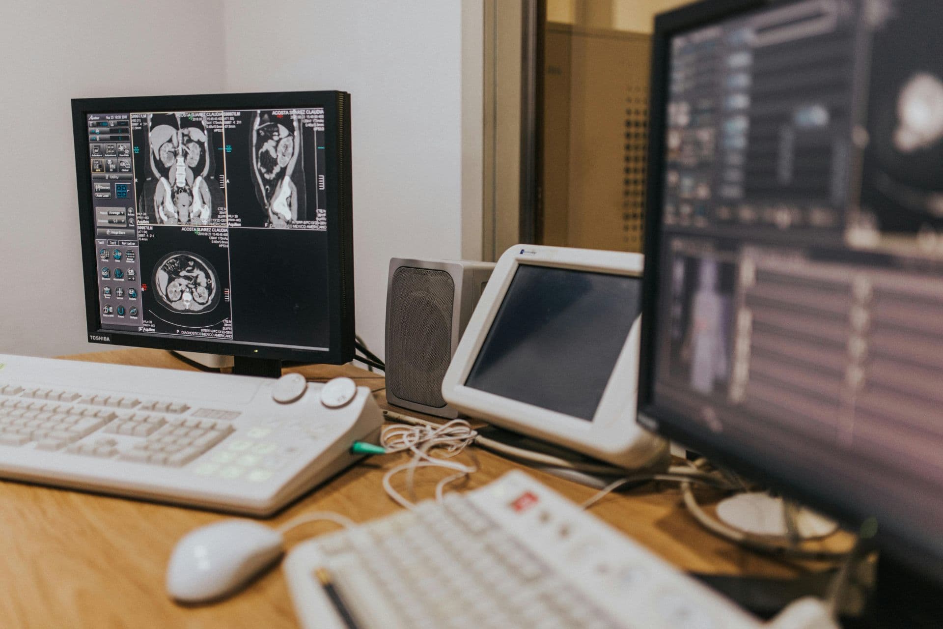
Radiology is the branch of medicine that uses imaging technology to view, diagnose, and sometimes treat diseases. It’s not just one machine; it includes X-rays, CT scans, MRI, ultrasound, mammography, and fluoroscopy.

Each tool works differently:
X-rays: Use small amounts of radiation to create images of bones and dense tissues. They are fast, affordable, and commonly used.
CT Scans: Combine hundreds of X-ray images from different angles to build detailed 3D pictures. They are especially useful for detecting tumors, injuries, or internal bleeding.
MRI: Uses strong magnets and radio waves not radiation to produce detailed images of soft tissues like the brain, muscles, and organs.
Ultrasound: Uses sound waves and is considered very safe. It is commonly used during pregnancy and for examining organs and blood flow.
One important thing many people misunderstand is radiation safety. Modern imaging machines are carefully designed to use the smallest safe dose possible. In radiography training, patient and staff protection is treated very seriously because safety always comes first.
Why Radiology Matters So Much
Radiology does far more than simply “take pictures.” In many cases, imaging is where treatment decisions begin. For example:
Mammograms can detect breast cancer early, increasing the chances of successful treatment.
CT scans can help doctors identify strokes within minutes, allowing faster emergency care.
Ultrasound helps guide biopsies and certain surgical procedures safely and accurately.
Without imaging, many conditions would be much harder to diagnose correctly. Radiology helps doctors move from uncertainty to clarity, which can make a life-saving difference for patients.
The Rise of AI and Smart Diagnosis
One of the biggest changes happening in radiology today is the use of Artificial Intelligence (AI).
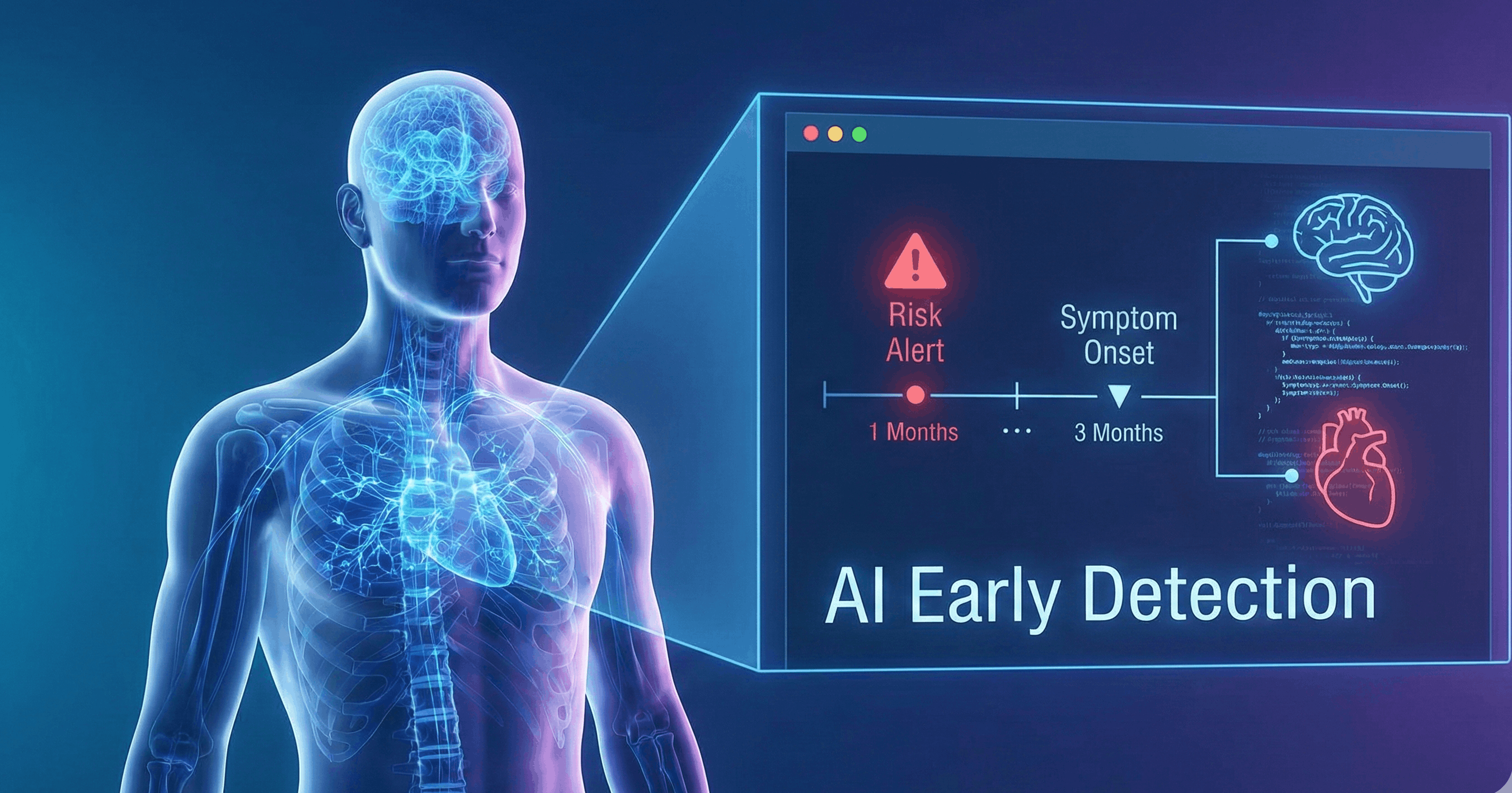
AI systems are trained using thousands or even millions of medical images. They can identify patterns, detect tiny abnormalities, and help prioritize urgent cases faster than before. In some situations, AI can spot small signs of disease that may be difficult to notice immediately.
But despite the excitement around AI, it is important to understand that it does not replace radiologists or radiographers. Instead, it works like a highly advanced assistant.
Healthcare professionals still analyze the images, make decisions, and communicate with patients. AI simply helps improve speed, efficiency, and accuracy.
For patients, this could mean: faster results, earlier diagnosis, fewer delays, and more effective treatment.
Conclusion
From Röntgen’s first X-ray in 1895 to today’s AI-assisted imaging systems, radiology has transformed healthcare in remarkable ways. What began as a groundbreaking discovery has become an essential part of modern medicine.
As a radiography student, it’s exciting to see how quickly the field continues to evolve. New technologies are making scans safer, more accurate, and more accessible to people around the world. Radiology is not just about machines or images. It is about seeing clearly, diagnosing correctly, and helping patients receive the care they need at the right time.


Discussion