Introduction
Every year, millions of people wait for organ transplants that may never come. Others live with chronic tissue damage from heart attacks, traumatic injuries, or degenerative diseases, where the body simply cannot repair itself fast enough.
Now imagine a different future: instead of waiting for a donor, doctors can implant lab-grown tissues designed specifically for your body. Instead of guessing how a drug might affect the liver or heart, researchers can test new drugs on engineered human tissue before it ever reaches patients.
This is the promise of tissue engineering. Tissue engineering is a field at the intersection of biology and engineering that aims to rebuild, repair, or replicate human tissues. And it’s not science fiction. Skin grafts grown in labs are already being used to treat burn victims. Engineered tissues are already helping scientists study cancer treatments, scarring, and cardiovascular disease in controlled settings.
So how does tissue engineering actually work? Let’s take a closer look.
The Core Components of Tissue Engineering
At its foundation, tissue engineering relies on three essential elements:
Cells
Structural Support
Biochemical and physical signals
Cells: The Living Workforce
Cells are the active builders of new tissues. Scientists may use stem cells, which can become many different types of cells, or more specialized cells, depending on the goal. These cells grow, organize, and produce their own support system. The support system is a natural material called the extracellular matrix that gives a tissue its strength and structure.
But cells need the right environment to do their job.
Structural Support: With or Without a Scaffold
Many engineered tissues use a scaffold, which is a supportive framework that gives cells structure and stability as they grow. Thanks to advances in biomaterials and 3D bioprinting, researchers can fine-tune scaffolds to control stiffness, shape, and porosity.
Why does that matter? Because cells respond to their surroundings. A soft surface can encourage stem cells to become one type of tissue, while a stiffer surface may guide them in a different direction.
Some scaffolds stay in place long term. Others are designed to gradually dissolve as natural tissue forms. If a scaffold breaks down too slowly, some cells might not function normally. If a scaffold breaks down too fast, the tissue may collapse on itself before it can function on its own. It’s a careful balance.
Other researchers skip scaffolds altogether. In scaffold-free approaches, cells cluster together and build their own support system. This method avoids potential issues like immune reactions, but can take longer and makes building large, strong tissues more challenging.
Signals: Chemical and Physical Instructions
Traditionally, scientists have focused on biochemical signals, such as growth factors, to guide cell behavior. But over the past two decades, researchers have realized that physical forces are just as important.
Cells don’t simply respond to chemistry. They respond to mechanics, too.
Why Mechanical Forces Matter
Inside the human body, cells are constantly experiencing physical forces. Heart muscles contract rhythmically. Lungs expand and recoil with every breath. Blood vessels feel the shear stress of flowing blood. Bone adapts to compression during movement.
These forces influence how cells grow, organize, and function through a process called mechanotransduction.
Mechanical cues can regulate:
Gene expression
Protein expression
Cell alignment
Extracellular matrix formation
Tissue maturation
If engineered tissues are meant to function like tissues in the body, they must experience something similar to the mechanical environment found in the body. Otherwise, they may look correct under a microscope but fail to fully function in real-world conditions.
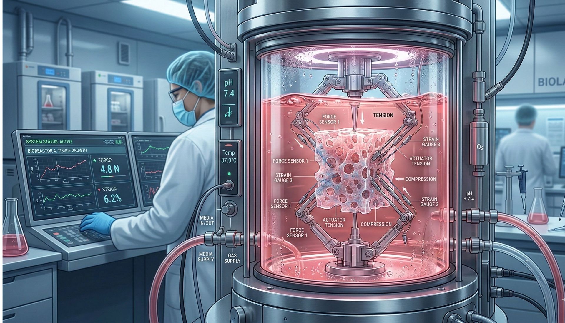
Bioreactors: Recreating the Body in the Lab
To deliver precise biophysical stimulation, scientists use devices known as bioreactors. These systems allow researchers to simulate the body’s mechanical environment in a controlled and reproducible way, ensuring each engineered tissue gets the same treatment.
Mechanical conditioning of engineered tissues can improve structural organization and enhance tissue function. It also enables scientists to investigate fundamental biological questions: How much force is needed to promote healthy growth? How much mechanical loading does it take to cause damage? How do mechanical and chemical signals interact?
By controlling physical forces in the lab, researchers can both engineer better tissues and deepen our understanding of human biology.
Beyond Regeneration: Modeling Disease
Tissue engineering isn’t limited to replacing damaged tissue. Engineered tissues are increasingly used to model disease processes, including scarring, cardiovascular remodeling, and cancer progression.
Because abnormal mechanical forces contribute to many diseases, recreating these conditions in the lab allows researchers to study cause-and-effect relationships more precisely than in the human body.
Additionally, modeling diseases allows researchers to study different therapeutics on tissues that more closely resemble the disease state found in humans.
This dual role, therapeutic and investigative, makes tissue engineering a powerful platform for both regenerative medicine and biomedical discovery.
Conclusion
Tissue engineering works by combining living cells, structural design, and carefully controlled biochemical and biophysical signals to recreate functional human tissue.
The field has evolved beyond simply placing cells onto materials. It now recognizes that cells are highly sensitive to their physical environment. Forces such as tension, compression, and fluid flow are not secondary details but rather essential biological instructors.
As researchers refine biomaterials, improve mechanical conditioning systems, and better understand how cells interpret physical cues, tissue engineering moves closer to its ultimate goal: creating tissues that not only survive, but truly function in the lab and inside the body.


Discussion