
The human ear is often described as one of the most sophisticated sensory systems in biology. Within a structure no larger than a seashell, the auditory system can detect frequencies ranging from the faint rumble of distant thunder to the subtle harmonic texture of a violin string. For decades, scientists believed they largely understood how this process worked. Sound waves entered the cochlea, traveled through fluid-filled chambers, and mechanically displaced the Basilar Membrane (BM), the ribbon-like structure that separates frequencies along its length much like keys on a piano.
Yet recent research has revealed that this classical model was incomplete.A 2026 study by Liu et al. published in the Journal of Anatomy uncovered an unexpected layer of biological complexity inside the human cochlea. The findings suggest that our extraordinary sensitivity to speech and music may depend not only on the mechanical properties of the Basilar Membrane itself, but also on a highly specialized elastic microarchitecture that actively shapes how sound is processed.
Rather than functioning as a passive acoustic surface, the cochlea appears to behave more like a living, continuously maintained biomechanical instrument.
The Active Architects: Why the Apex is the Ear’s Most Delicate Hardware
For many years, the Tympanic Covering Layer (TCL) was considered little more than a protective lining attached to the Basilar Membrane. Histological studies often treated it as a structural coating with limited physiological importance. However, high-resolution Transmission Electron Microscopy dramatically changed this interpretation.
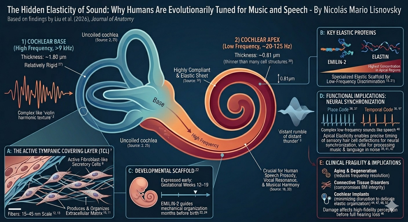
The cells composing the TCL were found to behave like active fibroblast-like secretory cells. Instead of remaining biologically inert, they continuously produce and organize extracellular matrix components that shape the membrane’s mechanical behavior. These cells deposit an amorphous ground substance alongside intricate fibrillary networks that form an elastic scaffold within the cochlea.
At the microscopic scale, the architecture is astonishingly delicate. Fibers measuring only 15–40 nanometers intertwine with broader fibrillar structures approximately 40–45 nanometers wide. Such dimensions approach the scale of molecular assemblies rather than conventional tissue anatomy.
This matters because hearing is fundamentally a problem of mechanical precision. Every spoken word, every musical tone, and every fluctuation in pitch depends on the controlled deformation of tissue structures thinner than many cellular membranes. The TCL therefore acts less like a passive covering and more like a biological engineering system responsible for maintaining the elasticity required for fine acoustic discrimination.
The effect is especially pronounced in the cochlear apex, the region specialized for low-frequency hearing. This apical zone is critically important for processing human speech prosody, vocal resonance, and musical harmony. The study (Liu et al., 2026) proposes that the active remodeling capacity of TCL cells may be one of the key evolutionary adaptations that gave humans unusually refined low-frequency auditory resolution.
The Protein Secret: EMILIN-2 and the Scaffolding of Development
One of the most important discoveries of the study was the identification of EMILIN-2 and elastin as major structural components of the human Basilar Membrane.
EMILIN-2, short for elastin microfibril interface-located protein 2, was previously (Russell et al., 2020) associated primarily with connective tissue organization in animal models. Its confirmed presence in the human cochlea suggests that hearing depends on a far more sophisticated elastic framework than previously recognized.
These proteins are not evenly distributed throughout the cochlea. Their concentration becomes particularly significant in the apical regions responsible for detecting low-frequency sound. In essence, the apex appears to be built with a specialized elastic composition optimized for slow, highly controlled vibrations.
The implications extend beyond adult hearing. RNA sequencing data demonstrated that both EMILIN-2 and elastin are expressed remarkably early during fetal development, between gestational weeks 12 and 19. This timing is critical because the cochlea undergoes rapid morphological remodeling during this developmental window.
As the fetal ear expands and coils into its mature form, the extracellular matrix must remain sufficiently elastic to preserve structural integrity while simultaneously guiding precise anatomical organization. EMILIN-2 likely functions as a developmental scaffold, ensuring that the Basilar Membrane acquires the exact biomechanical properties necessary for mature auditory function.
This finding reinforces an important principle in developmental biology: function is often embedded into tissues long before the organism begins using them. The machinery for music and language may therefore begin assembling months before birth.
A Tale of Two Coils: Mapping Elasticity Through the Cochlea
The cochlea is not mechanically uniform. Its physical properties change dramatically from base to apex, creating a frequency map along its spiral length. Using Greenwood’s formula alongside synchrotron radiation phase-contrast imaging, researchers quantified these regional differences with unprecedented detail. The basal cochlea, responsible for detecting high-frequency sounds above roughly 9 kHz, contains a relatively thick and rigid Basilar Membrane measuring approximately 1.80 micrometers in thickness. Structurally, this region resembles the cochleae of many laboratory mammals. The apex, however, is radically different.
In the low-frequency regions corresponding to approximately 20–125 Hz, the membrane becomes extraordinarily thin, approaching only 0.81 micrometers. This is thinner than many single-cell structures and nearly an order of magnitude smaller than the diameter of a human red blood cell. Such extreme thinness creates a highly compliant mechanical environment capable of responding to very subtle low-frequency vibrations. The membrane here behaves less like a rigid platform and more like a finely tensioned elastic sheet.
This specialization may explain why humans possess unusually advanced low-frequency discrimination compared to many mammals. Human speech relies heavily on low-frequency temporal cues, including intonation, rhythm, emotional inflection, and harmonic structure. Music similarly depends on the precise interpretation of resonance and timing within these lower acoustic ranges. The cochlear apex therefore appears to represent a uniquely human adaptation optimized for complex auditory communication.
The Timing Advantage: How Humans Resolve the Voice
Traditional auditory theory emphasized what is known as a “place code,” meaning that different frequencies activate different physical regions of the Basilar Membrane. While this principle remains valid, the new findings suggest that human low-frequency hearing may depend equally on timing. Instead of relying solely on where a vibration occurs, the auditory system may also depend on when neural signals fire relative to one another. The elastic properties of the Basilar Membrane influence the velocity and synchronization of sensory hair cell deflections, allowing auditory neurons to preserve temporal precision.
This synchronization is essential for separating meaningful sounds from noisy environments. The human brain can isolate a familiar voice in a crowded room because auditory neurons maintain exquisitely coordinated firing patterns that preserve timing information within milliseconds.
The study proposes that the specialized elastic constituents found in the apical cochlea help sustain this synchronization. In other words, the elasticity of the membrane itself may contribute directly to the neural coding of speech and music. Human auditory evolution may therefore have favored not only sharper frequency tuning, but also greater temporal fidelity.
The Vulnerability Factor: When the Machinery of Sound Fails
Understanding the cochlea’s elastic microarchitecture also reveals how fragile the auditory system truly is. If hearing depends on nanometer-scale fiber organization and highly specialized extracellular proteins, even subtle structural disruptions may impair auditory resolution long before complete hearing loss occurs. Damage does not necessarily have to destroy the cochlea outright; it may simply interfere with the fine mechanical synchronization required for high-fidelity sound perception.
This has major implications for aging and disease. Connective tissue disorders and autoimmune diseases capable of altering glycoprotein organization may directly compromise the structural integrity of the Basilar Membrane. Likewise, age-related degeneration could progressively disrupt the organization of elastic fibers, reducing frequency discrimination and temporal precision over time.
The findings may also reshape how scientists think about cochlear implants. Although modern implants can restore remarkable levels of hearing, electrode insertion introduces comparatively large foreign structures into an extraordinarily delicate biomechanical environment. Even minor disruption of the membrane’s elastic organization could alter the very frequency resolution the implant seeks to recover.
The study further highlights the importance of spiral ganglion neurons in the apical cochlea. These neurons cluster within confined anatomical spaces where ephaptic interactions, subtle electrical influences between neighboring neurons, may contribute to signal synchronization. Disturbing this organization could impair timing-dependent aspects of auditory perception even when the sensory cells themselves remain functional.
A New Frontier in Auditory Science
The emerging picture of the human cochlea is far more dynamic than previously imagined. Hearing is not simply the passive vibration of a membrane struck by sound waves. It is an actively maintained biomechanical process involving secretory cells, elastic proteins, nanoscale fiber networks, and exquisitely synchronized neural timing.
By identifying the fibroblast-like behavior of Tympanic Covering Layer cells and clarifying the structural role of EMILIN-2, this research opens new possibilities for auditory medicine. Future cochlear implants, regenerative therapies, and treatments for hearing loss may need to preserve or recreate not only sensory cells, but also the delicate extracellular architecture that enables human acoustic precision.
Our capacity to perceive the warmth of a familiar voice, the emotional depth of music, or the subtle cadence of spoken language may ultimately depend on biological structures only a few hundred nanometers thick. The more we uncover about the cochlea, the clearer it becomes that human hearing is not merely sensitive — it is exquisitely engineered, fragile, and deeply tied to the evolutionary history of communication itself.



Discussion